Microscope Experiment Idea: The Food We Eat
What’s more interesting to a child (and more useful to teach as a parent!) than about the foods we ingest? Although what you’re able to view may differ depending on what kind of microscope you have, there’s plenty to put under a microscope and show sitting right in your pantry and refrigerator! This type of microscope experiment will help differentiate what different kinds of foods are made of (such as plant cells of a slide of onion versus bacterial cells of yogurt), and keeps costs down since almost everything is right there in your home.
– Yogurt Microscope Experiment

Recommended Microscope Options:
- AmScope B120C (40x-2500x, LED lit compound microscope)
- Omax CS-M82ES-SC100-LP50 (40x-2000x, LED lit compound microscope w/ slides package)
Recommended Equipment:
- Glass Microscope Slides & Cover Slips
- Dropper
- Yogurt with live culture (Activia or Yakult are common in grocery stores)
- Methylene Blue Stain (Gram Stain Kit)
- Paper Towels
Microscope Experiment Process:
- Take a very small drop of yogurt using the dropper, and place it on the center of the glass slide. Using a cover slip, gently flatten the yogurt drop.
- Place a drop of methylene blue next to the edge of the cover slip. Use a paper towel on the opposite side of the cover slip to draw some yogurt out, which will draw some of the methylene blue into the sample.
- Note: Methylene blue is optional in this microscope experiment, as, the yogurt is visible without it, however, the stain helps with additional contrast. Stain will kill off the bacteria, so using it without stain will allow you to see more excited bacteria.
- Place the slide on the microscope stage, with the sample centered on the aperture (hole) of the stage, where the light is focused on the sample. Adjust the diaphragm (the iris
- View in the compound microscope at 4x to start, and focus by adjusting the focusing knobs on the microscope until the image comes into clear view. Move your way up to the 10x, 40x, and eventually 100x objectives (be sure to use immersion oil with the 100x objective, as we outlined in our Microscope Education section). Bacteria are very small, and will appear small even at higher objective magnifications, however they are clearly visible.
- Study what you see in the microscope. The bacteria should be shaped in different clusters, either isolated, in pairs, or threads, and can be shaped like rods or spheres. Pairs denoted by containing “diplo” in the name, threads “strepto,” and rods are “bacilli,” while spheres are coccus.
- Additional research note: How is yogurt made?
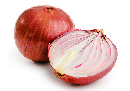
– Onion Microscope Experiment

Recommended Microscope Options:
- AmScope B120C (40x-2500x, LED lit compound microscope)
- Omax CS-M82ES-SC100-LP50 (40x-2000x, LED lit compound microscope w/ slides package
Recommended Equipment:
- Onion
- Scalpel
- Forceps (Tweezers)
- Dropper
- Glass Microscope Slides & Cover Slips
- Water
- Iodine
- Paper Towels
Microscope Experiment Process:
- Cut thin layer of onion or onion skin, small enough to fit on a side and as thin as possible.
- Place the piece of onion on the glass slide with a small drop of water to keep it from wilting or drying out.
- Place the cover slip over the sample.
- Drop a drop of iodine next to the cover slip, and use the paper towel on the opposite side to drawn in some water, which will draw the iodine into the sample.
- Place the slide on the microscope stage, with the sample centered on the aperture (hole) of the stage, where the light is focused on the sample. Adjust the diaphragm (the iris
- View in the compound microscope at 4x to start, and focus by adjusting the focusing knobs on the microscope until the image comes into clear view. Move your way up to the 10x, 40x, and eventually 100x objectives (be sure to use immersion oil with the 100x objective, as we outlined in our Microscope Education section).
- Since onion cells are plant cells, try to identify as many structures in the cell as you can see. Additional research note for this experiment is to draw an image of a plant cell and label the cell structures and their functions.
– Lettuce/Spinach Microscope Experiment

Recommended Microscope Options:
- AmScope B120C (40x-2500x, LED lit compound microscope)
- Omax CS-M82ES-SC100-LP50 (40x-2000x, LED lit compound microscope w/ slides package
Recommended Equipment:
- Lettuce or Spinach
- Scalpel
- Forceps (Tweezers)
- Dropper
- Glass Microscope Slides & Cover Slips
- Water
- Iodine
- Paper Towels
Microscope Experiment Process:
- Cut thin square of either leafy green as available, small enough to fit on a side and as thin as possible.
- Place the piece on the glass slide with a small drop of water to keep it from wilting or drying out.
- Place the cover slip over the sample.
- Stain or die is not needed for this microscope experiment, due to the natural color the leafy green has.
- Place the slide on the microscope stage, with the sample centered on the aperture (hole) of the stage, where the light is focused on the sample. Adjust the diaphragm (the iris
- View in the compound microscope at 4x to start, and focus by adjusting the focusing knobs on the microscope until the image comes into clear view. Move your way up to the 10x, 40x, and eventually 100x objectives (be sure to use immersion oil with the 100x objective, as we outlined in our Microscope Education section).
- Since lettuce and spinach cells are both plant cells, try to identify as many structures in the cell as you can see. Additional research note for this experiment is to draw an image of a plant cell and label the cell structures and their functions, if it hasn’t been done for the onion experiment.
- If it has, additional research ideas would be to compare and contrast the difference between an onion cell and a leafy green cell.
– Bread Mold Microscope Experiment

Recommended Microscope Options:
- AmScope B120C (40x-2500x, LED lit compound microscope)
- Omax CS-M82ES-SC100-LP50 (40x-2000x, LED lit compound microscope w/ slides package
Recommended Equipment:
- Molded Bread
- Glass Microscope Slides & Cover Slips
- Methylene Blue Stain
- Disposable Gloves
- Face Mask
- Dropper
- Water
- Paper Towels
Microscope Experiment Process:
- Take a slice of bread and set it aside. Allow the bread to sprout mold, however, be sure to use gloves and potentially a face mask to avoid contamination by breathing mold spores in. For this experiment, please leave about two weeks for the bread to mold (therefore, start this with enough time to complete the experiment before any due dates!)
- Once bread has molded, place a drop of water on the center of the glass slide. Scrape a little of the mold into the water, and cover with a cover slip.
- Place a drop of methylene blue stain on one side of the cover slip on the glass slide, then on the opposite side, use a paper towel to absorb some water, drawing the methylene blue into the sample and staining your mold spores. This improves contrast in your sample, greatly helping visibility for this microscope experiment.
- View in the compound microscope at 4x to start, and focus by adjusting the focusing knobs on the microscope until the image comes into clear view. Move your way up to the 10x, 40x, and eventually 100x objectives (be sure to use immersion oil with the 100x objective, as we outlined in our Microscope Education section).
- For additional research ideas, try to look online and figure out different types of mold that can grow on bread, and for extra credit, try to identify which one grew on your bread!
Stay tuned for more ideas! Any questions, comments, concerns, or other microscope experiment ideas, please email or leave a comment! 🙂
